Daisy framework projects
24th January, 2011A collaborative effort.
A collaborative effort.
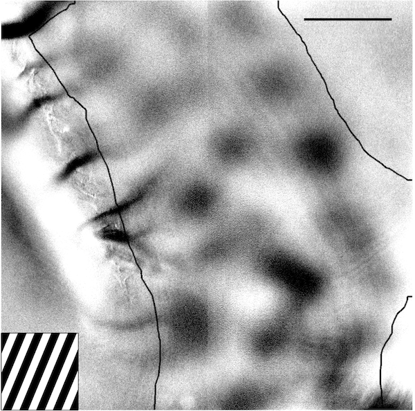
When injections of dye are made into the upper layers of cortex, a remarkable structure of neuronal connectivity is revealed. Dye is absorbed by neurons and transported along axons and dendrites to fill entire cells. When a large population of neurons is stained in this way, slices examined in tangential section show a semi-regular array of denser staining.
This pattern is remarkable simply because it appears in every area of cortex, over a wide range of animals. The pattern, reminiscent of the petals of a daisy, and the yet unknown connectivity system which underlies it, is a general feature of the cortical anatomy. Since spatially segregated areas of cortex perform radically different functions (from visual processing to movement control to higher cognition), this connectivity system could form part of a general, adaptable computation engine in cortex.
Analysis of the structure of OI response maps
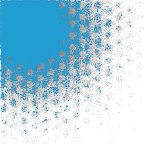
Neurons in the visual cortex respond to the visual world with an intricate pattern of activity. Locations across the surface of the cortex respond strongly to some visual inputs, and weakly to others. This can be seen in the figure at left, which shows a "map" of how a small area of visual cortex responds to a picture of several black and white bars. These maps show that neurons that are active for a particular visual input are grouped into small regions of cortex (several of which can be seen in the figure).
The roughly periodic projections of the superficial patch system are evocative of the rough periodicity present in cortical maps. In this project we examined to what extent the structure of this anatomical system (the patch system) constrains the structure of the functional system (maps of the cortical response). We developed techniques for automatically locating the clusters of active neurons in a functional map, and comparing their spatial arrangement with that of the superficial patch system.
We found that the spatial rules used to lay out the patch system mirror the rules used to lay out responsive regions across the surface of primary visual cortex. The structure of the patch system, and of cortical maps, is also highly conserved across species.
See the project summary for more information. This work was published in Cerebral Cortex.
Investigating the structure of the superficial patch system
What is the geometric structure of the individual axonal projections that collectively form the superficial patch system? We explored whether patch system-like patterns could be reproduced in a model of cortical projections. The complex labelling patterns produced by labelling in primary visual cortex (area 17) defy most simple explanations. We showed that a combination of several geometric rules, consistent with reasonable expectations of primary visual cortex structure and function, were sufficient to produce the complexities of patch system labelling patterns in area 17.
See the project summary for more information. This work was published in Cerebral Cortex.
Funding
These projects were supported by the European Commission [FP6 2005-015803 DAISY]; and by the John Crampton Travelling Scholarship.